
Scheuermann disease Radiology Case Radiology, Disease, Thoracic vertebrae
Scheuermann disease is an osteochondrosis that causes localized changes in vertebral bodies, leading to backache and kyphosis. Diagnosis is with spinal x-rays. Treatment usually involves only reduction of weight bearing and strenuous activity. Scheuermann disease manifests in adolescence and is slightly more common among boys.

Image
Introduction. Scheuermann's disease (SD), also known as Scheuermann's juvenile kyphosis or juvenile osteochondrosis of the spine, was first described by Holger Scheuermann in 1920. SD develops prior to the onset of puberty, typically presenting during the adolescent growth spurt as a rigid, sometimes painful thoracic kyphosis. 1.

Scheuermann disease Radiology Case
Lumbar Scheuermann disease is a type of variant Scheuermann disease where there is no abnormal kyphosis. This has been reported in the lumbar spine and thoracolumbar junction of patients of all ages, and back pain may be present. On imaging, affected individuals can have vertebral endplate changes, disc space narrowing, and anterior Schmorl.

Dr Balaji Anvekar FRCR Scheuermann's disease MRI
Scheuermann's disease is a self-limiting skeletal disorder of childhood. Scheuermann's disease describes a condition where the vertebrae grow unevenly with respect to the sagittal plane; that is, the posterior angle is often greater than the anterior. This uneven growth results in the signature "wedging" shape of the vertebrae, causing kyphosis.
Hyperkyphosis Scheuermanns Disease
Aim: To find accompanying anomalies of typical and atypical Scheuermann's disease (SD) is reported in the present study. Methods: Study included 20 patients (16 men and 4 women) who had radiological imaging radiography, magnetic resonance imaging (MRI) and computed tomography, if available, due to back pain, curved back and low back pain in November 2011-February 2016 period.

Scheuermann's Kyphosis Spine Orthobullets
Therefore, in the present study, we aimed to evaluate the radiological pathomorphological features of the spine and the association between the thoracic kyphosis angle and the clinical presentation by assessing pain and self-perceived body image in patients with Scheuermann's disease. We proposed two hypotheses.

Scheuermann's disease Radiology Case
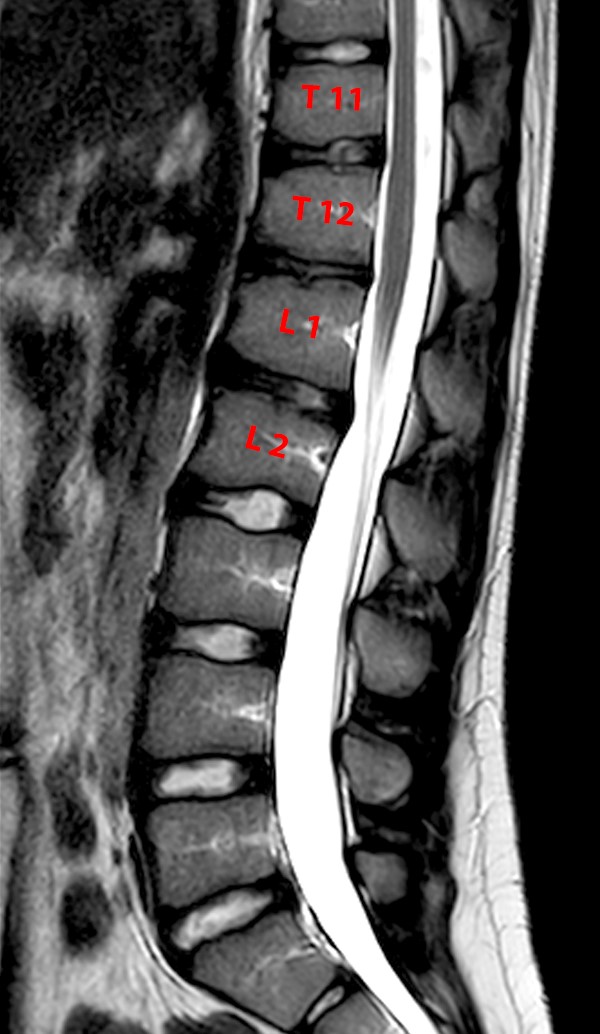
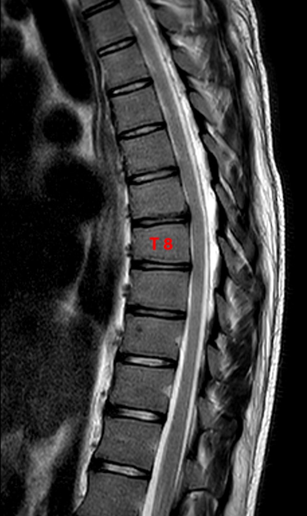
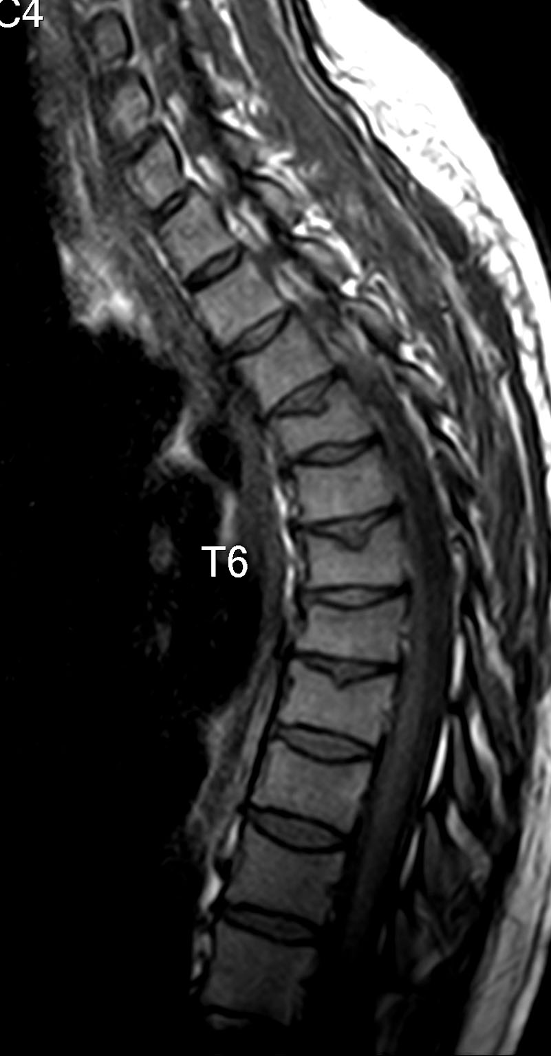
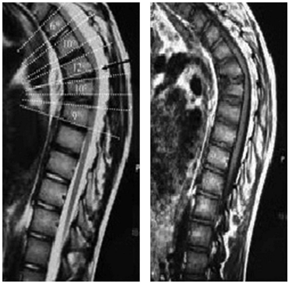
STIR. Coronal. STIR. Axial. T2. Decreased lower thoracic and lumbar vertebral height, associated with marked irregularity of end-plates and formation of multiple prominent Schmorl's nodes. The AP diameter of the spinal canal gradually decreases as the level lowers.
Scheuermann’s Disease Radsource
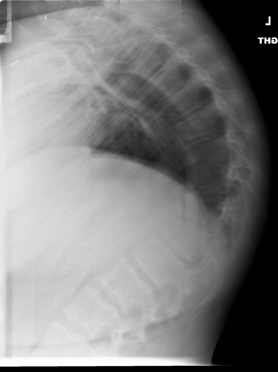
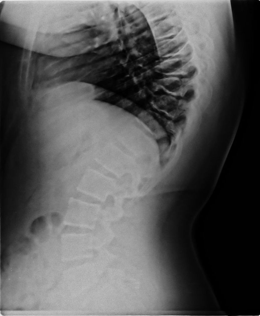
Sagittal. bone window. Axial bone. window. There is mild, chronic wedging of lower thoracic vertebral bodies, with Schmorl's nodes and disc space narrowing. No acute compression fracture. Facet joints are enlocated. Posterior elements are intact. The appearance of the thoracic spine is consistent with Scheuermann's disease.
Scheuermann’s Disease Radsource
Scheuermann disease, (juvenile kyphosis), is a growth disturbance with curving deformity of the thoracic or thoracolumbar spine in adolescents that causes an increase bowing or rounding of the back in the sagittal plane. It is defined by anterior vertebral wedging of at least 5° in 3 or more adjacent vertebral bodies.
MY ERADIOLOGY CASES December 2010
Scheuermann's Kyphosis (SK), first described by Horgel Welfer Scheuermann in 1920, is a rigid spinal kyphosis usually involving the thoracic or thoracolumbar area ( 1 ). Albeit several theories, its etiology is still unknown ( 2 ). SK is usually separated into two groups: typical and atypical SK. Typical SK is the more common type and has a mid.
scheuermanndisease4 PinkyBone
Scheuermann's disease (SD) is a progressive disease associated with back pain or low back pain in adolescents. It is the most common cause of structural thoracic or thoracolumbar hyperkyphosis, and could be seen in typical or atypical patterns. Diagnosis of SD is made by radiological methods along with clinical findings.
Scheuermann’s Disease POGO Physio Gold Coast
The theories relating to Scheuermann's disease are reviewed. An attempt is made to resolve discrepancies in these theories in the light of radiologically derived evidence. This evidence suggests that central nodes, marginal nodes, and the irregular ossification of Scheuermann's disease, result from different mechanisms, and that the problem has been obscured by failure to consider them as.

Scheuermann disease Image
Scheuermann Disease / therapy*. Thoracic Vertebrae / diagnostic imaging. Recent studies have revealed a major genetic contribution (a dominant autosomal inheritance pattern with high penetrance and variable expressivity) to the etiology of Scheuermann kyphosis with a smaller environmental component (most probably mechanical factors).

Scheuermann disease presenting as compressive myelopathy Neurology
Notably, both classic SD and atypical SD are associated with back pain. 14, 16, 17, 19 - 22 Although Scheuermann kyphosis is uncommon, SD radiological signs have been observed in 18% to 40% of the general population, 23 suggesting that SD, or more precisely, "SD-like" spine, may be a variant of normal spine morphology rather than a.
Scoliosis and Kyphosis, what's the difference? Scoliosis Clinic UK Treating Scoliosis
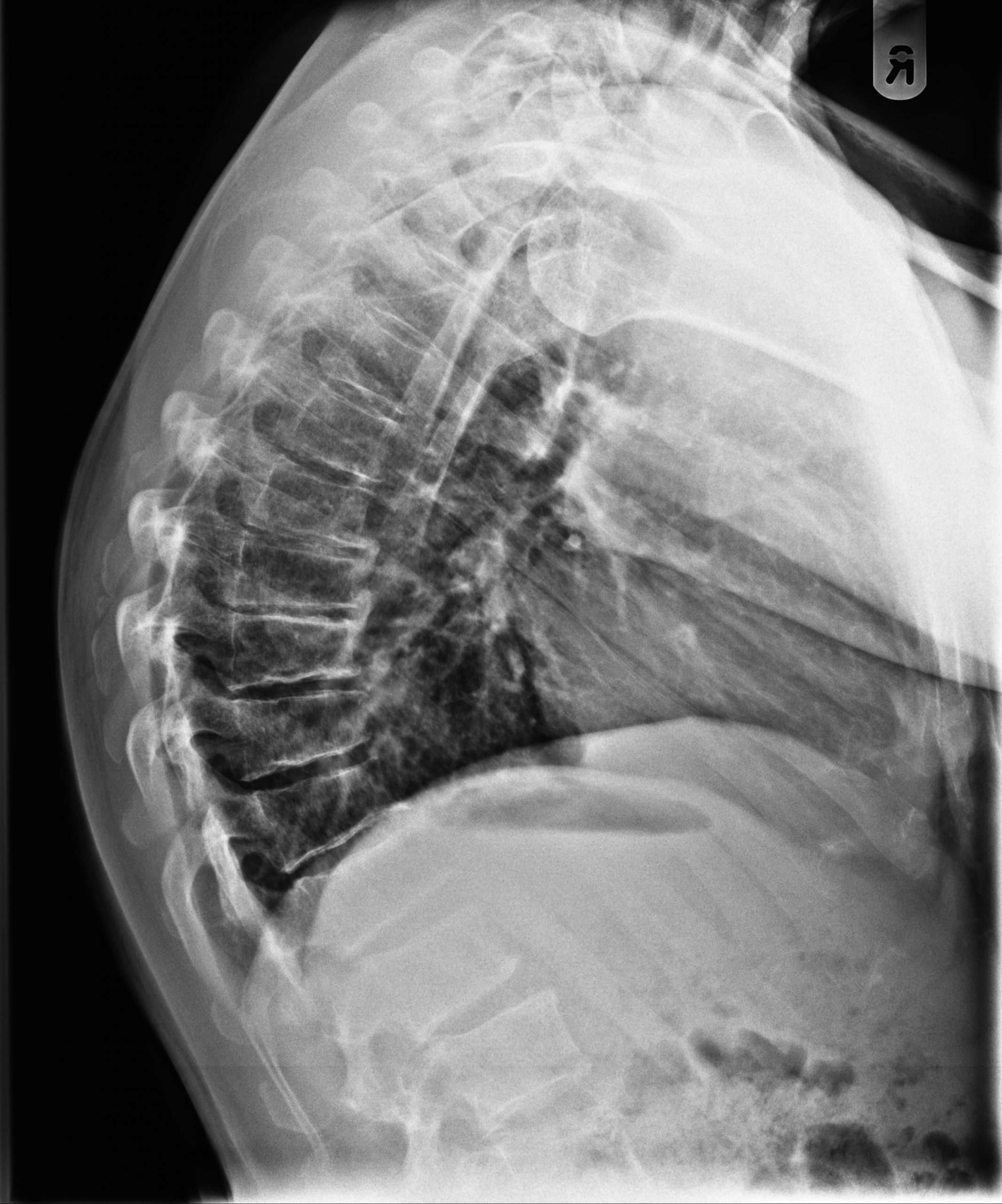
Gender: Female. x-ray. Vertebral wedging, subchondral osseous irregularity in the endplates, Schmorl's nodes and an asymptomatic thin cleft in the neural arch of the fifth lumbar vertebra. There's also hypoplasia of the left iliac bone and mild hypoplasia of the ipsilateral proximal femur.

Morbus Scheuermann Gesundheitsportal Medavit De My XXX Hot Girl
Case Discussion. A rare example of Scheuermann disease "caught in the act", that may eventually result in kyphosis. Also note the persisting neurocentral synchondroses typical for that age.